Watch the delivery of our MRI building, a key step in bringing advanced imaging closer to home for our community.
At Mason District Hospital, our Imaging Department provides a full range of diagnostic services in a compassionate, convenient setting—close to home.
We combine state-of-the-art technology with highly trained, caring technologists,
offering truly High-Tech, High-Touch care. Our team guides you every step of the way,
making your experience as comfortable and reassuring as possible.
For questions or to schedule a service call (309) 543-8596.
Our Services
- Computed Tomography (CT) Imaging
- Dual-Energy X-Ray Absorptiometry (DEXA)
- Magnetic Resonance Imaging (MRI)+
- Mammography
- Nuclear Medicine
- Positron Emission Tomography – Computed Tomography (PET-CT)
- Ultrasound
- X-Ray
Computed Tomography (CT) Imaging
- What it is: CT scans use X-rays and computers to produce detailed cross-sectional
images of the body.
- What to expect: You will lie on a padded table that moves through the scanner. The procedure is fast and designed to minimize discomfort, even for children and seniors.
- Why it’s important: Provides precise images to help your doctor diagnose conditions accurately and efficiently.
- Who performs it: Certified technologists guide you through every step for a smooth, reassuring visit.
All of these services are offered through using state-of-the-art equipment and are provided by highly-trained, professional technologists. Another way Mason District Hospital is High-Tech, High-Touch.
Dual-Energy X-Ray Absorptiometry (DEXA)
- What it is: Measures bone density to detect osteoporosis and assess fracture risk.
- What to expect: You will lie on a padded table for about 25 minutes. Safe, painless, no special preparation, and no clothing changes required.
- Why it’s important: Helps your doctor evaluate bone health and develop a plan to reduce fracture risk.
- Who is at risk: Family history of osteoporosis, low body weight, low calcium intake, smoking, mobility issues, hyperthyroidism, age.
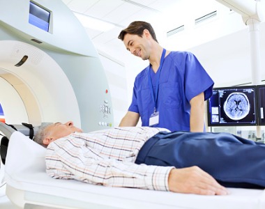
Magnetic Resonance Imaging (MRI)
- What it is: MRI uses magnets, radio waves, and computers to create detailed images
of organs, tissues, and bones.
- What to expect:
- You will lie on a padded table that slides into a large magnet.
- Non-invasive, no radiation exposure.
- Who performs MRI exams?
MRI exams are performed by certified MRI technologists using Mason District Hospital’s onsite Canon 1.5 Tesla MRI system. The images are then reviewed and interpreted by board-certified radiologists, ensuring accurate and timely results for your healthcare provider.
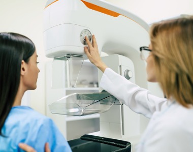
Mammography
- What it is: Digital Mammography uses low-dose X-rays to create images for early
detection of breast abnormalities.
- What to expect:
- The scan is quick and performed by a certified technologist.
- Images are shared immediately with your physician for faster results.
- When to start: Annual screening is recommended starting at age 40 by leading health organizations.
- Who performs it: Certified mammographers committed to patient comfort and accurate results.
Nuclear Medicine
- What it is: Uses a very small amount of radioactive material to examine organ function
and detect abnormalities.
- Types of exams:
- Single Photon Emission Computed Tomography (SPECT)
- Whole-body scans
- Cardiac imaging with stress tests
- Thyroid imaging and treatment
- Why it’s important: Assesses kidney function, heart blood flow, lung health, gallbladder, bones, and detects cancer.
- Who performs it: Certified nuclear technologists dedicated to patient comfort and care.
Positron Emission Tomography – Computed Tomography (PET-CT)
- What it is: Combines PET (shows body function) and CT (shows body structure) for a
complete diagnostic view.
- What to expect:
- The PET tracer may be injected, swallowed, or inhaled depending on the exam.
- The CT scan produces detailed images to locate abnormalities.
- Why it’s important:
- Detect and monitor cancer
- Evaluate brain disorders such as Alzheimer’s, epilepsy, or tumors
- Assess heart disease and blood flow
- Who performs it: Mobile imaging services with a GE Discovery integrated PET and CT scanner in one gantry.
Ultrasound
- What it is: Ultrasound (sonography) uses high-frequency sound waves to create
images of your body.
- What to expect:
- A gel is applied to the skin and a handheld probe is moved across the area.
- Non-invasive, painless, and safe for adults, children, and babies.
- Why it’s important: Provides real-time images for diagnosing a variety of conditions.
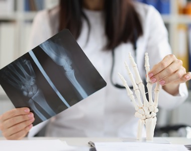
X-Ray
- What it is: X-rays use a focused beam of photons to produce images of bones and soft
tissues.
- What to expect:
- Quick, painless imaging.
- Some exams may require specific positioning, guided by a technologist.
- Why it’s important: Commonly used to detect broken bones and other conditions.
- Who performs it: Highly trained technologists ensuring safe, accurate imaging.
Radiology Interpretation
All exams are reviewed by OnRad, Inc. Radiology Services, a team of board-
certified radiologists available 24/7, 365 days a year.
They focus on accuracy, reliability, and timely results to support your doctor in providing
the best care.